|
|
Epidermoid Cyst of the Testicle
Submitted by Matt Bokermann, MD
General Considerations
- Rare testicular lesion accounting for 1-2% of testicular tumors
- Benign testicular tumor with no malignant potential
- Most commonly, this lesion occurs in the 2nd-4th decades of life
Clinical findings
- Clinical presentation usually involves testicular enlargement or a palpable mass without other clinical symptoms
- More common in the right testicle. There have been very rare cases of bilateral epidermoid cysts.
Ultrasound Imaging findings
- Ultrasound is the study of choice
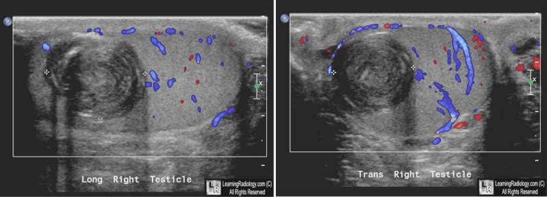
- Ultrasound will show a round and well demarcated intratesticular mass
- Internal heterogeneity, often with alternating hyperechoic and hypoechoic internal rings with an “onion skin” appearance.
- A hyperechoic center is often present.
- May have a hyperechoic or hypoechoic rim
- Avascular on color imaging
MRI Imaging findings
- Well demarcated intratesticular mass.
- T2 weighted imaging shows high signal intensity that may have low intensity internal foci and a low intensity rim
- Absence of enhancement with contrast
Pathologic correlation
- Thought to represent monodermal (i.e., only ectodermal) formation of a teratoma.
- May also involve squamous metaplasia of the seminiferous epithelium or rete testes.
- Echogenic rim corresponds to a fibrous capsule made of squamous epithelium
- Layering “onion skin” appearance corresponds with layering of keratin and squamous cells that have undergone metaplasia
Treatment
- Making a pre-surgical diagnosis is important, as it may allow testicular-preserving surgery rather than orchiectomy
- Testicular-preserving surgery is usually satisfactory when frozen sections show an epidermoid cyst and two biopsies of the surrounding testicular parenchyma show no neoplasm


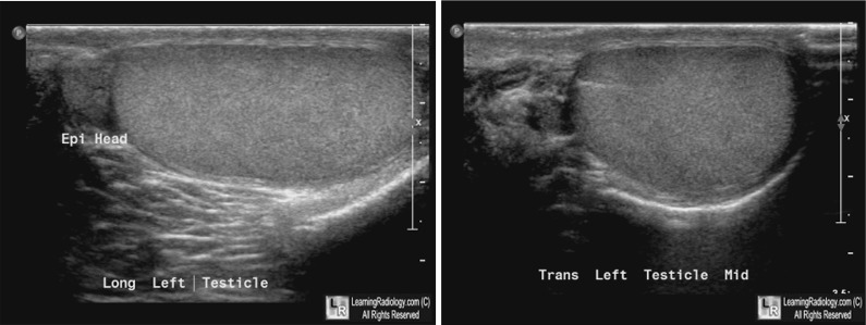
Epidermoid Cyst of the Right Testicle. Longitudinal and transverse images of the right testicle (above)
show a mass with concentric echoic and hypoechoic rings pathognomonic
for an epidermoid cyst of the testicle (white arrows) The normal left testicle is shown below.
For these same photos without the arrows, click here and here
For more information, click on the link if you see this icon 
Epidermoid Cyst of the Testis: Radiologic-Pathologic Correlation. Alma G Loya et al; RadioGraphics, 2004, S243-S246
Sonographic and MR Imaging Findings of Testicular Epidermoid Cysts.Cho et al;American Journal Roentgenology 2002; 178:743-748
|
|
|
